Advertisement
Advertisement
October 2026
Sponsored by Medtronic
Carotid Artery Stenting With Integrated Embolic Protection for High-Risk Symptomatic Internal Carotid Artery Stenosis

Carotid endarterectomy and carotid artery stenting are established revascularization options for patients with carotid artery stenosis.1-3 Selection of the revascularization approach should account for medical conditions, lesion anatomy, surgical accessibility, and procedural risk.2,3 In particular, a high cervical lesion (a distal lesion located above the level of the second cervical vertebra) may be difficult to access surgically and can increase the complexity of carotid endarterectomy.4,5 Recent neurologic events, including stroke, may further increase the procedural risk during the carotid endarterectomy, particularly when anesthesia is required.3,4 In such patients, carotid artery stenting may provide an alternative treatment strategy.
Embolization during carotid artery stenting remains an important procedural concern, particularly during stent deployment and postdilatation.6,7 The Neuroguard IEP™* 3-in-1 Carotid Stent and Post-Dilatation Balloon System with Integrated Embolic Protection (Contego Medical, Inc.) combines a closed-cell carotid stent, integrated postdilatation balloon, and 40-µm integrated filter.7,8 The system is a rapid exchange platform and should always be used with a commercially available primary distal embolic protection device as described in the Instructions for Use.7,8 The PERFORMANCE II trial evaluated the Neuroguard IEP System in 305 patients at elevated risk for adverse events associated with carotid endarterectomy and reported a procedural success rate of 96.7%, with no major strokes through 30 days.8
The case highlighted below describes transfemoral carotid artery stenting in a symptomatic patient with a severe, surgically inaccessible lesion in the high cervical left internal carotid artery (ICA).
CASE PRESENTATION
Patient Presentation
A woman in her 70s with a known history of hypercoagulable state (treated with apixaban) had an acute ischemic stroke manifested by right-sided weakness. She received intravenous thrombolytic therapy followed by neurointervention with thrombectomy of the left anterior communicating artery and left ICA, with improvement in her motor deficit. After 3 days of hospitalization, she was transferred to an acute rehabilitation facility, where her neurologic function continued to improve. Despite the initial improvement, she continued to experience intermittent neurologic symptoms, including diplopia, blurred vision, and slurred speech.
Diagnostic Evaluation and Treatment Decision
Further evaluation demonstrated severe residual stenosis of the left ICA. Because the lesion was located high above the carotid bifurcation, the patient was referred for catheter-based angiography and consideration of endovascular intervention. About 1 month after the acute stroke, carotid angiography demonstrated an approximate 95% stenosis of the distal left ICA at a high cervical location (Figure 1A). Cerebral angiography also showed impaired intracranial perfusion, with absent or markedly reduced filling of the anterior cerebral artery (Figure 1B). Because the lesion was located high above the carotid bifurcation, making the lesion difficult to access surgically, carotid endarterectomy was considered unsuitable. Her recent stroke was also considered to increase the risk associated with anesthesia. Given the severe symptomatic stenosis, ongoing intermittent symptoms, and unfavorable anatomy for endarterectomy, transfemoral carotid artery stenting was selected.
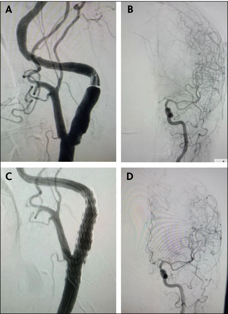
Figure 1. A preintervention carotid angiogram demonstrating an approximate 95% stenosis of the high cervical left ICA (A). A preintervention cerebral angiogram showing impaired intracranial perfusion and reduced or absent filling of the anterior cerebral artery (B). A completion carotid angiogram demonstrating restoration of luminal diameter and improved flow through the treated left ICA (C). A post-intervention cerebral angiogram showing improved intracranial perfusion, including restoration of anterior cerebral artery filling (D).
Intervention
A Spider™ FX distal embolic protection filter (Medtronic) was advanced across the lesion and deployed in the distal ICA. The Neuroguard IEP System was then delivered across the stenosis. After deployment of the 40-µm integrated embolic protection (IEP) filter, the carotid stent was deployed across the lesion. The integrated balloon was used for postdilatation, after which the 40-µm integrated filter was collapsed and the delivery system was removed. Completion angiography demonstrated a widely patent treated segment with substantially improved flow through the left ICA (Figure 1C). Intracranial angiography showed restoration of cerebral perfusion, including renewed filling of the anterior cerebral artery (Figure 1D).
Outcome
The patient experienced immediate symptomatic improvement after revascularization. No new neurologic deficit was reported during the postprocedural hospitalization, and she was discharged home the following day. At 1-week follow-up, the patient reported complete resolution of her speech deficits and upper extremity weakness and had returned to work. Residual right lower extremity weakness persisted but continued to show improvement.
Discussion
This case demonstrates successful transfemoral carotid artery stenting in a patient with recurrent neurologic symptoms and an approximate 95% stenosis of a high cervical ICA, considered high risk for carotid endarterectomy. Utilization of the Neuroguard IEP System in conjunction with a primary distal embolic protection filter resulted in an excellent angiographic result at the target stenosis and restored normal intracranial perfusion. The patient had an immediate improvement in symptoms and no new postprocedural neurologic deficit.
Although this case involved symptomatic carotid stenosis, the recently reported CREST-2 data provide important context for the management of asymptomatic high-grade carotid stenosis. Among asymptomatic patients receiving intensive medical therapy for high-grade carotid stenosis (≥ 70%), the addition of carotid artery stenting reduced stroke risk, whereas carotid endarterectomy did not provide a significant benefit over medical therapy alone.9 This evidence, combined with the availability of next-generation carotid stent systems, supports the consideration of carotid artery stenting in appropriately selected patients.
In my experience with > 300 cases using the Neuroguard IEP System, successful delivery of the stent system was achieved in 99% of procedures. No immediate strokes were observed, while three groin access complications occurred. Although these single-operator observations are not a substitute for prospective comparative evidence, they do provide additional real-world support for the device’s deliverability and immediate procedural safety when used in appropriately selected patients. Careful preprocedural imaging and planning remain essential. Lesion morphology, distal landing zone, vessel diameter, arch anatomy, tortuosity, calcification, antiplatelet eligibility, and the ability to safely deliver both primary embolic protection and IEP should be evaluated before intervention.
CONCLUSION
Transfemoral carotid artery stenting with the Neuroguard IEP System and its 40-µm filter, used in conjunction with primary distal embolic protection, achieved successful revascularization of a symptomatic, approximate 95% high cervical left ICA stenosis. Restoration of intracranial perfusion was accompanied by immediate symptomatic improvement, with discharge the following day and return to work the next week. This case illustrates the utility of carotid artery stenting in appropriately selected symptomatic patients with high cervical lesions that may be technically challenging for surgical revascularization.
1. Brott TG, Hobson RW II, Howard G, et al. Stenting versus endarterectomy for treatment of carotid-artery stenosis. N Engl J Med. 2010;363:11-23. doi: 10.1056/NEJMoa0912321
2. Müller MD, Lyrer P, Brown MM, Bonati LH. Carotid artery stenting versus endarterectomy for treatment of carotid artery stenosis. Cochrane Database Syst Rev. 2020;2:CD000515. doi: 10.1002/14651858.CD000515.pub5
3. Bonati LH, Kakkos S, Berkefeld J, et al. European Stroke Organisation guideline on endarterectomy and stenting for carotid artery stenosis. Eur Stroke J. 2021;6:I-XLVII. doi: 10.1177/23969873211012121
4. White CJ, Brott TG, Gray WA, et al. Carotid artery stenting: JACC state-of-the-art review. J Am Coll Cardiol. 2022;80:155-170. doi: 10.1016/j.jacc.2022.05.007
5. White CJ, Ramee SR, Collins TJ, et al. Carotid artery stenting: patient, lesion, and procedural characteristics that increase procedural complications. Catheter Cardiovasc Interv. 2013;82:715-726. doi: 10.1002/ccd.24984
6. Obeid T, Arnaoutakis DJ, Arhuidese I, et al. Poststent ballooning is associated with increased periprocedural stroke and death rate in carotid artery stenting. J Vasc Surg. 2015;62:616-623.e1. doi: 10.1016/j.jvs.2015.03.069
7. Langhoff R, Schofer J, Scheinert D, et al. Double filtration during carotid artery stenting using a novel post-dilation balloon with integrated embolic protection. JACC Cardiovasc Interv. 2019;12:395-403. doi: 10.1016/j.jcin.2018.11.039
8. Gray WA, Metzger DC, Zidar J, et al. The PERFORMANCE II trial: a prospective multicenter investigation of a novel carotid stent system. JACC Cardiovasc Interv. 2025;18:367-376. doi: 10.1016/j.jcin.2024.10.031
9. Brott TG, Howard G, Lal BK, et al. Medical management and revascularization for asymptomatic carotid stenosis. N Engl J Med. 2026;394:219-231. doi: 10.1056/NEJMoa2508800
Disclosures
Dr. Attanti: Consultant for Medtronic.
This material should not be considered the exclusive source of information, it does not replace or supersede information contained in the device manual(s). Please note that the intended use of a product may vary depending on geographical approvals. See the device manual(s) for detailed information regarding the intended use, the procedure, indications, contraindications, warnings, precautions, and potential adverse events. Medtronic products placed on markets comply with legislation (if applicable) on medical devices.
If you are located in the United States, please refer to the brief statement(s) below to review applicable indications, safety and warning information. For further information, contact your local Medtronic representative and/or consult the Medtronic website at www.medtronic.com.
Neuroguard IEP™* 3-in-1 Carotid Stent and Post-Dilatation Balloon System with Integrated Embolic Protection
Brief Statement
Indications for Use
The Neuroguard IEP™* 3-in-1 Carotid Stent and Post-Dilatation Balloon System with Integrated Embolic Protection is indicated for improving the carotid luminal diameter in subjects at high risk for adverse events from carotid endarterectomy who require carotid revascularization and meet the criteria outlined below:
- Patients with symptomatic stenosis of the common or internal carotid artery with ≥ 50% as determined by angiography using NASCET methodology, OR Patients with asymptomatic stenosis of the common or internal carotid artery with ≥ 80% as determined by angiography using NASCET methodology.
- Patients with reference vessel diameters 4.0 – 8.0 mm.
This device is also indicated for post-dilation of the stent component with simultaneous capture and removal of embolic material. The Neuroguard IEP System should always be used in conjunction with an available primary distal embolic protection device as described in the IFU.
Contraindications
The Neuroguard IEP™* 3-in-1 Carotid Stent and Post-Dilation Balloon System with Integrated Embolic Protection is contraindicated for use in:
- Patients in whom anticoagulant and/or antiplatelet therapy is contraindicated;
- Patients with a known hypersensitivity to nickel-titanium;
- Patients with severe vascular tortuosity or anatomy that would preclude the safe introduction of a guidewire, catheter, introducer sheath, delivery system or embolic protection device;
- Patients with uncorrected bleeding disorders;
- Patients with known hypersensitivity to heparin, including those patients who have had a previous incident of Heparin-Induced Thrombocytopenia (HIT) type II.
Warnings
- The appropriate antiplatelet, anticoagulant and if necessary, vasodilator therapy, must be used during the pre- and post-procedure to minimize the risk of embolism and thrombus.
- Maintain continuous flush while inserting and removing the Neuroguard IEP System over the guidewire. Perform all exchanges slowly to prevent air embolism or trauma to the artery.
- Never withdraw or move an intravascular device against any resistance until the cause is determined. Applying excessive force during delivery or retrieval of the Neuroguard IEP System can potentially result in loss or damage to the device or components or injury (perforation/dissection) to the vessel.
Multiple stents should not be used in an overlapping configuration.
- A partially deployed stent cannot be repositioned or retracted into the outer sheath. Retraction, or repositioning of a stent by force, could cause deformation of the stent, damage to the blood vessel and/or to the delivery catheter. The stent cannot be removed after implantation.
- Balloon inflation pressure should not exceed the labeled rated burst pressure (RBP). Exceeding the rated burst pressure (RBP) may result in balloon rupture, which can traumatize the artery.
- The stent may cause thrombus, distal embolization or may migrate from the site of implant down the arterial lumen. Appropriate sizing of the stent to the vessel is required to reduce the possibility of stent migration. In the event of thrombosis of the expanded stent, thrombolysis and PTA should be attempted.
- This device contains nitinol, an alloy of nickel and titanium. Persons with allergic reactions to these metals may suffer an allergic reaction to this implant. Prior to implantation, patients should be counseled on the materials.
- The Neuroguard IEP System should always be used with a primary distal embolic filter. The device was studied with the FilterWire™ EZ, Emboshield® NAV6, and SpiderFX™ filters. The safety and effectiveness has not been evaluated with other embolic protection systems.
Precautions
- Special care must be taken not to disrupt the stent or the filter on the delivery system.
- Avoid over-tightening the hemostasis valve as this may affect balloon inflation/deflation times as well as movement of the guidewire.
- If resistance is met during delivery system introduction, the system should be withdrawn and another system used.
- Only prepare and flush the Neuroguard IEP System with the supplied flushing syringes and flushing tips.
- Venous access should be available during carotid stenting to manage bradycardia and/or hypotension by either pharmaceutical intervention or placement of a temporary pacemaker, if needed.
- Use only recommended balloon inflation solution. Do not use air or other gaseous medium to inflate the balloon. Nonionic contrast medium has different viscosity and precipitation levels than does the ionic type, which may prolong inflation/deflation times.
- The delivery system is not designed for use with power injection. Use of power injection may adversely affect device performance.
- Caution should be used if pre-dilating the lesion without embolic protection as this may increase the risk of an adverse outcome.
- Do not advance or retract the Neuroguard IEP System unless the balloon is fully deflated under vacuum and the filter is fully collapsed. If resistance is met during manipulation, determine the cause of the resistance before proceeding.
Potential Adverse Effects
Potential complications include, but are not limited to the following: angina, allergic reactions (including to antiplatelet agents, contrast medium or stent materials), aneurysm, arrhythmias, arterial occlusion/thrombosis at puncture site, bleeding from anticoagulant or antiplatelet medications, bradycardia, carotid artery spasm, cerebral edema, cerebral hemorrhage, cerebral ischemia/transient ischemia attack (TIA), cardiac tamponade, cardiogenic shock, death, detachment and/or implantation of a component, embolism, fever, filter thrombosis/occlusion, groin hematoma with or without surgical repair, heart failure, hematoma, hemorrhage, hypotension/hypertension, infection, ischemia/infarction of tissue/organ, myocardial infarction, pain and tenderness, pericardial effusion, pulmonary edema, pseudoaneurysm at the vascular access site, renal failure/insufficiency, respiratory failure, restenosis of the stented segment, seizure, severe unilateral headache, stent embolization, stent/filter entanglement/damage, stent malapposition, stent migration, stent misplacement, stent thrombosis/occlusion, stroke/cerebrovascular accident (CVA), total occlusion of carotid artery, vessel dissection, perforation, spasm or recoil, vessel trauma requiring surgical repair or reintervention.
CAUTION: Federal (USA) law restricts this device to sale by or on the order of a physician.
Important information: Indications, contraindications, warnings and instructions for use can be found in the product labeling supplied with each device, or contact a Medtronic representative.
SpiderFX™ embolic protection device
Reference Statement
Indications for Use
Lower Extremity (LE) Interventions
The SpiderFX™ Embolic Protection Device is indicated for use as a guidewire and embolic protection system to contain and remove embolic material in conjunction with the TurboHawk™ Peripheral Plaque Excision System, either during standalone procedures or together with PTA and/or stenting, in the treatment of severely calcified lesions in arteries of the lower extremities. The vessel diameter at the filter basket placement site should be between 3.0mm and 6.0mm.
Carotid Interventions
The SpiderFX™ Embolic Protection Device is indicated for use as a guidewire and embolic protection system to contain and remove embolic material (thrombus/debris) while performing angioplasty and stenting procedures in carotid arteries. The diameter of the artery at the site of filter basket placement should be between 3.0mm and 7.0mm.
Saphenous Vein Graft (SVG) Interventions
The SpiderFX™ Embolic Protection Device is indicated for use as an embolic protection system to contain and remove embolic material (thrombus/debris). The device also acts as the guidewire while performing percutaneous transluminal coronary angioplasty or stenting procedures in coronary saphenous vein bypass grafts with reference vessel diameters of 3.0mm to 6.0mm. The safety and effectiveness of this device as an embolic protection system has not been established in the cerebral vasculature.
Contraindications
Do not use with patients in whom anticoagulant and antiplatelet therapy is contraindicated; Do not use in vessels with excessive tortuosity; Do not use in patients with uncorrected bleeding disorders; Do not use in the renal arteries.
Potential Adverse Events
The potential complications include, but are not limited to the following: Access site adverse event (e.g., av fistula, hematoma, hemorrhage, pseudoaneurysm, puncture site infection); Adjunct device entanglement (LE only); Adverse reaction to antiplatelet/anticoagulation agents or contrast media; Allergic reaction to device materials; Amaurosis Fugax (carotid only); Amputation (LE only); Aneurysm (LE and SVG); Angina (carotid and SVG only); Arterial dissection; Cerebral hemorrhage (carotid only); Congestive heart failure (carotid only); Cardiac tamponade (SVG only); Coronary ischemia (SVG only); Death; Device(s) deformation, collapse, fracture, or rupture; Device(s) thrombosis (acute and subacute); Dissection (LE and carotid only); Embolization of air, debris, plaque or thrombus (evidenced by slow or no-flow) from mechanical disruption by the intervention resulting in TIA or stroke; Embolization or migration of the interventional device(s); Emergency surgery; Failure to deliver stent to the intended site (SVG only); GI bleeding due to anticoagulation (LE and carotid only); Hemodynamic compromise (e.g., prolonged hypotension requiring treatment with intravenous medications) (LE and carotid only); Hyperperfusion syndrome (carotid only); Hypotension or hypertension; Infection; Intimal flap (SVG only); Intracerebral bleed (carotid only); Ischemia (LE and SVG only); Myocardial infarction; Occlusion (SVG only); Renal insufficiency (LE and carotid only); Renal failure requiring dialysis; Repeat intervention to treatment site (LE only); Restenosis of stented segment (greater than 50% obstruction) (SVG only); Seizure (carotid only); Sepsis; Significant cardiac arrhythmia requiring treatment with medications and/or transvenous pacing; Stent entanglement; Stroke (LE and carotid only); Thrombosis (acute and subacute); Transient Ischemia Attack (TIA) (LE and carotid only); Vasospasm; Vessel dissection, perforation, rupture or intimal flap; Vessel/filter occlusion
CAUTION: Federal (USA) law restricts this device to sale by or on the order of a physician.
Important information: Indications, contraindications, warnings and instructions for use can be found in the product labeling supplied with each device, at www.medtronic.com/manuals or contact a Medtronic representative
US-CV-2602385 ©2026 Medtronic. Medtronic is the sole authorized distributor of commercially available Contego Medical products in the United States. Medtronic, Medtronic logo, are trademarks of Medtronic. All other brands are trademarks of a Medtronic company. Please refer to product labeling for indications, contraindications, warnings, and instructions for use. Contact your local Medtronic representative for availability or more information. For Global distribution. 10/2026
The Neuroguard IEP® System 3-in-1 Carotid Stent and Post-Dilation Balloon System with Integrated Embolic Protection is not approved for distribution outside of the United States.
Advertisement
Advertisement