Advertisement
Advertisement
September 2026
Sponsored by Becton, Dickinson and Company
From Long-Segment Occlusions to ISR: The Rotarex™ Catheter System
Two experienced operators discuss the versatility of the Rotarex™ Catheter System for treating complex peripheral artery disease (PAD), with case examples highlighting its use in long-segment occlusions and in-stent restenosis (ISR).


First, can you tell us about your practice and the peripheral artery disease (PAD) patients you treat?
Dr. Foteh: I am the Chief of Vascular Surgery at Vital Heart and Vein, a multispecialty cardiovascular practice in Houston, Texas. Our organization includes 6 vascular surgeons and more than 30 cardiologists, representing a broad range of specialties, including interventional cardiology and electrophysiology. We also have 3 podiatrists on staff, as we do a lot of limb salvage and wound care.
We treat a broad spectrum of PAD patients, from those with intermittent claudication to individuals with nonhealing wounds and chronic limb-threatening ischemia. Limb salvage is a major focus of our practice, and we manage PAD across the full spectrum of disease severity.
Dr. Joye: I’ve been a practicing interventional cardiologist for almost 30 years now and have been deeply invested in the vascular care of my patients since the very beginning of my career. Previously, I spent a lot of time in device development when treatment options were far more limited than today. I’ve tried just about everything out there to try to get the best results for my patients. I continue to do a high volume of vascular cases on an annual basis, working in surgical centers and hospitals in Northern California.
What differentiates the Rotarex™ Catheter System from other treatment options that you consider?
Dr. Foteh: The Rotarex™ Catheter System is very versatile. It can treat acute thrombus, chronic thrombus and mixed-etiology plaques like fibro thrombotic plaques. Its ability to address multiple lesion types makes it my go-to device when it comes to atherectomy interventions.
Dr. Joye: For me, the Rotarex™ Catheter System offers two major benefits. First, the setup is fast, straightforward and easy for both me and my staff to manage. And most importantly, in my experience, when the device is used appropriately, I have found that slow flow and downstream debris tend to occur infrequently compared with other treatment options.
CASE SPOTLIGHT: Addressing Long-Segment Native Vessel ISR Occlusions With the Rotarex™ Catheter System
By Kousta Foteh, M.D.
CASE PRESENTATION
A man in his early 70s presented with a several-month history of lifestyle-limiting claudication after walking approximately 150 to 300 ft. The patient was an ex-smoker with a history of hypertension, coronary artery disease and peripheral vascular disease. He had undergone intervention 8 years prior to presentation for claudication, with placement of a stent in his superficial femoral artery (SFA). A duplex ultrasound examination revealed an occluded SFA and an ankle-brachial index (ABI) of 0.61. We initiated medical therapy that included antiplatelet therapy as well as cilostazol and asked him to participate in a walking program. The patient returned 3 months later following treatment, and there was no improvement in his walking distance. It was decided at that time to proceed with reintervention.
PROCEDURAL OVERVIEW
A pedal approach was used to obtain vascular access. Retrograde angiography revealed a long-segment SFA occlusion extending from just above the patella to the SFA ostium, consisting of mixed calcified and thrombotic plaque.1 Atherectomy was performed with the Rotarex™ Catheter System, which was advanced through the diseased SFA segment. Follow-up angiography revealed favorable luminal gain following effective debulking, aspiration and atherectomy. A 6 X 300 mm Lutonix™ 018 drug-coated balloon (DCB) was used as adjunctive therapy, and final angiography showed complete resolution of the occlusion with no residual stenosis or dissection and preserved two-vessel runoff.2
POST PROCEDURE FOLLOW-UP
This patient recovered well after the procedure and was walking the next day. At 1- and 6-month follow-up, a post procedure duplex ultrasound showed a widely patent SFA with no signs of stenosis and an ABI of 0.94. The patient continues to do well with no claudication.
DISCUSSION
This case highlights the versatility of the Rotarex™ Catheter System in treating both ISR and long-segment native vessel occlusions. Its effectiveness across moderately calcified, thrombotic and mixed plaque, combined with the ability to use it from multiple access approaches, makes the Rotarex™ Catheter System a valuable front-line tool for the treatment of PAD.
1Do not use the device in calcified vessel segments that exhibit radiopacities on both sides of the arterial wall and extend beyond 10 mm in length prior to contrast injection or digital subtraction angiography.
2Vessel preparation using only pre-dilatation was studied in the LEVANT 2 clinical study. Other methods of vessel preparation, such as atherectomy, have not been studied clinically with the Lutonix™ DCB Catheter.
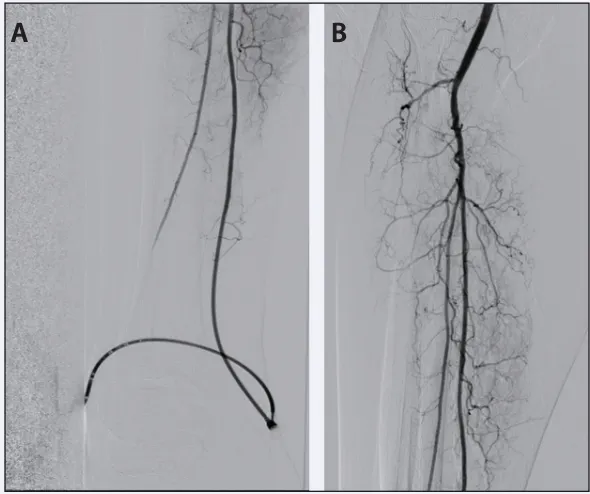
Figure 1. Lesion crossed using 0.018-inch Quick-Cross™ catheter (A). Successful intraluminal passage (B).
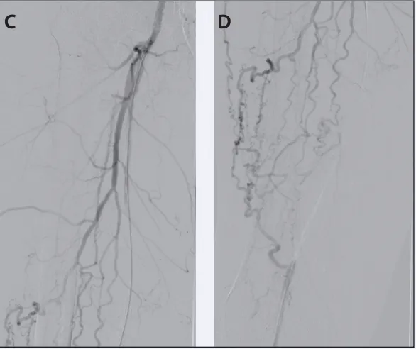
Figure 2. SFA occlusion (C, D).
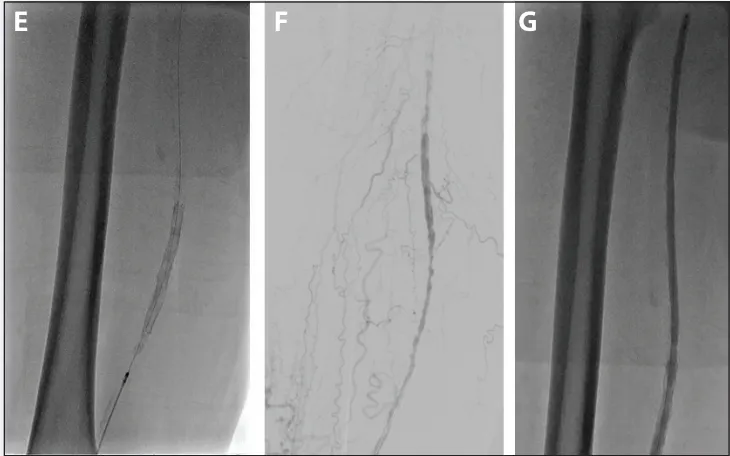
Figure 3. Atherectomy using the Rotarex™ Catheter System (E). Post atherectomy results (F). Adjunctive angioplasty performed using Lutonix™ 018 DCB (G).
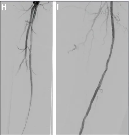
Figure 4. Final results (H, I).
How frequently do you use pedal access when performing procedures with the Rotarex™ Catheter System? Do you have recommended techniques or best practices for this approach?
Dr. Joye: I perform a fair amount of pedal access cases, and so this technology is attractive when disease extends into the distal popliteal artery. In these cases, preserving multiple tibial vessels is critical.
When you’re coming from an antegrade position, oftentimes you’re faced with a wire going into a single tibial vessel and you may wall off a seemingly viable other tibial vessel for runoff in the process. In my pedal cases where I use the Rotarex™ Catheter System, I’m using the pedal access to get retrograde through a lesion, which typically involves the bifurcation or trifurcation of the tibial arteries so that I can preserve as many tibial vessels as possible.
Once I’ve got my pedal access, I still favor going back into the true lumen upstream, doing a snare retrieval, reversing the wire, and treating it in an antegrade fashion. Generally, I use the pedal access for getting where I want to get to and then reversing the wire and utilizing Rotarex™ Catheter System in an antegrade fashion.
Dr. Foteh: Pedal access is my primary approach for a majority of my patients because in my opinion, it’s safer than retrograde femoral access as it minimizes complications like pseudoaneurysm, postoperative pain, and time to recuperation and walking. A few best practices can help ensure safe and effective use of the device. First, confirm the tibial vessel is at least 3 mm in diameter and that there are no tibial lesions that could prevent the device from passing safely. In some cases, adjunctive therapy may be required to optimize the tibial vessels before advancing the device.
If you encounter resistance while advancing the Rotarex™ Catheter System through a tibial vessel, avoid forcing it forward. Instead, withdraw the device and reassess the vessel, which may include repeat imaging to identify any underlying issue. If safe passage cannot be achieved, it is best to abandon atherectomy with the Rotarex™ Catheter System or, alternatively, get retrograde femoral access and treat from there.
CASE SPOTLIGHT: Restoring Flow in a Long-Segment SFA ISR Occlusion With the Rotarex™ Catheter System
By James D. Joye, D.O.
CASE PRESENTATION
A woman in her mid-80s with a history of hypertension, hyperlipidemia and diabetes mellitus presented with claudication. She reported always being active but suddenly started experiencing worsening symptoms, now only being able to walk a block or two before having to stop because of calf claudication. A duplex ultrasound was performed, which identified severe disease in her right SFA and proximal popliteal artery.
Initial angiography revealed long-segment disease from the proximal to mid-SFA extending to the proximal segment of the popliteal artery (P1). Although I would generally default to a percutaneous bypass in cases like this, the caliber of her vessels and first presentation with vascular disease led me to choose a more conventional treatment approach, consisting of pre-dilation with a PTA balloon, followed by stent placement.
REINTERVENTION
A few months later, the patient returned with recurrent, lifestyle-limiting claudication. Surveillance ultrasound suggested significant ISR, which was confirmed on angiography, demonstrating diffuse restenosis throughout the stented segment. Given the patient’s small-caliber, two-vessel tibial runoff, the Rotarex™ Catheter System was the clear treatment choice. Atherectomy was performed using both antegrade and pullback passes, followed by DCB angioplasty. Final angiography showed increased luminal gain, minimal residual stenosis and brisk tibial runoff with no evidence of distal embolization or other downstream sequelae.
DISCUSSION
This case highlights the challenge of treating long-segment femoropopliteal disease. Although conventional stenting initially provided symptom relief, the patient developed ISR within months. Treatment with the Rotarex™ Catheter System followed by DCB angioplasty achieved meaningful luminal gain and restored flow, demonstrating an effective approach for managing complex femoropopliteal ISR while avoiding additional stent placement.
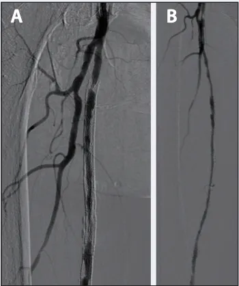
Figure 1. ISR of the proximal SFA (A) and right SFA/popliteal artery (B).
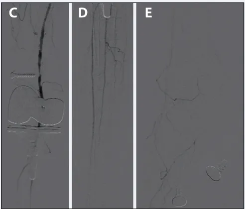
Figure 2. ISR in P1 and two-vessel runoff via the anterior/peroneal arteries (C), tibial runoff (D), and faint pedal runoff (E).
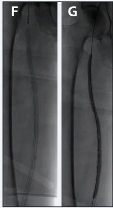
Figure 3. Rotarex™ Catheter System (F) and DCB (G).
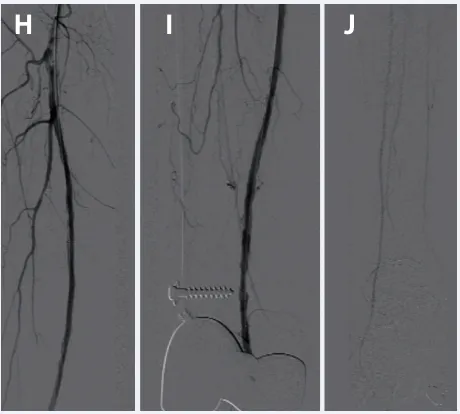
Figure 4. Final result (H-J).
When treating in-stent restenosis (ISR), what factors lead you to select Rotarex™ Catheter System over other treatment options?
Dr. Joye: One of the biggest changes in my practice has been adopting the Rotarex™ Catheter System as my first-line treatment for ISR. After using a variety of other technologies, I was often dissatisfied with either the limited lumen gain or inadequate aspiration. Today, I use the Rotarex™ Catheter System in more than 95% of my ISR cases because it is easy to set up, tracks through lesions well, and delivers the luminal gain I look for. Most importantly, it effectively removes and aspirates debris, which, in my experience, has significantly reduced the risk of downstream embolization.
Dr. Foteh: Its mechanism of action, which combines active aspiration with mechanical thrombectomy, represents a distinct advantage here. Not only does it effectively debulk plaque and thrombotic material within the stent, but it also removes that material during the procedure, resulting in luminal gain I seek to achieve.
Secondly, another major advantage is the confidence it gives me with respect to distal embolization. Because the device aspirates debris as it works, I am less concerned about clinically significant downstream embolization. For that reason, when I’m treating ISR or an in-stent occlusion, the Rotarex™ Catheter System is my go-to device every time.
Rotarex™ Atherectomy System
The Rotarex™ Atherectomy System is intended for use as an atherectomy device and to break up and remove thrombus from native peripheral arteries or peripheral arteries fitted with stents, stent grafts or native or artificial bypasses.
The Rotarex™ Atherectomy System is contraindicated in patients not suitable for atherectomy/thrombectomy; in the cardiopulmonary, coronary, carotid, cerebral and renal vasculature; in vessels that are undersized for the device used; and in the venous vasculature.
Potential adverse events include, but are not limited to: Embolization, especially distal embolization · Pulmonary embolisms of all degrees of severity · Thrombosis · Re-occlusion · Vessel wall injury · Vessel dissection/perforation/rupture · Perforation as a result of mural calcium being torn out of the vessel wall · Arteriovenous fistula/pseudo-aneurysm · Hematoma, bleeding, hemorrhage · Organ perforation · Implants such as stents/stent grafts/bypass grafts getting damaged, caught or dislodged · Disruption of the catheter: debris remaining in the body · Allergic reactions, including allergic reactions to device components · Infections or necrosis at the puncture site · Catheter-induced sepsis · Death.
Lutonix™ 018 Drug Coated Balloon
The Lutonix™ 018 Drug Coated Balloon PTA catheter is indicated for percutaneous transluminal angioplasty, after appropriate vessel preparation, of de novo, restenotic, or in-stent restenotic lesions up to 300 mm in length in native superficial femoral or popliteal arteries with reference vessel diameters of 4-7 mm. The Lutonix™ 018 Drug Coated Balloon PTA catheter is indicated for percutaneous transluminal angioplasty, after pre-dilatation, for treatment of stenotic lesions of dysfunctional native arteriovenous dialysis fistulae that are 4 mm to 7 mm in diameter and up to 80 mm in length. The Lutonix™ Catheter is contraindicated for use in: 1) Patients who cannot receive recommended anti-platelet and/or anticoagulant therapy. 2) Women who are breastfeeding, pregnant or are intending to become pregnant or men intending to father children. It is unknown whether paclitaxel will be excreted in human milk and there is a potential for adverse reaction in nursing infants from paclitaxel exposure. 3) Patients judged to have a lesion that prevents complete inflation of an angioplasty balloon or proper placement of the delivery system.
Potential adverse events which may be associated with a peripheral balloon dilatation procedure include: Additional intervention ∙Allergic reaction to drugs, excipients, or contrast medium ∙Amputation/loss of limb (SFA) ∙Aneurysm or pseudoaneurysm ∙Arrhythmias ∙Embolization ∙Hematoma ∙Hemorrhage, including bleeding at the puncture site ∙Hypotension/hypertension ∙Inflammation ∙Loss of permanent access (AVF) · Occlusion ∙Pain or tenderness ∙Pneumothorax or hemothorax (SFA) ∙Sepsis/infection ∙Shock ∙Steal Syndrome (AVF) · Stroke ∙Thrombosis ∙Vessel dissection, perforation, rupture, or spasm. Although systemic effects are not anticipated, refer to the Physicians’ Desk Reference for more information on the potential adverse events observed with paclitaxel. Potential adverse events, not described in the above source, which may be unique to the paclitaxel drug coating include: Allergic/immunologic reaction to the drug coating (paclitaxel) ∙Alopecia ∙Anemia ∙Blood product transfusion ∙Gastrointestinal symptoms ∙Hematologic dyscrasia (including leukopenia, neutropenia, thrombocytopenia) ∙Hepatic enzyme changes ∙Histologic changes in vessel wall, including inflammation, cellular damage, or necrosis ∙Myalgia/Arthralgia ∙Myelosuppression ∙Peripheral neuropathy.
Please consult respective product labels and instructions for use for indications, contraindications, hazards, warnings and precautions. Lutonix™ DCB is Rx only.
Disclaimers: This article is sponsored by Becton, Dickinson and Company (BD). Drs. Foteh and Joye are paid consultants of BD. The opinions and clinical experiences presented herein are for informational purposes only. The results from the case studies discussed herein may not be predictive for all patients. Individual results may vary depending on the variety of patient specific attributes.
BD, the BD Logo, Lutonix and Rotarex are trademarks of Becton, Dickinson and Company or its affiliates. All other trademarks are the property of their respective owners. © 2026 BD. All Rights Reserved.
BD-182984
Advertisement
Advertisement