Advertisement
Advertisement
October 2026 Supplement
Sponsored by Medtronic
Case Study: Extensive Echogenic DVT Managed With the Liberant™ Thrombectomy System

PATIENT PRESENTATION
A female patient in her mid-40s presented to the emergency department with acute-onset swelling and pain in her right leg persisting for the last 6 days. This was preceded by an upper respiratory infection the week before. She denied any chest pain, shortness of breath, or palpitations. A review of her medical history revealed prior diagnoses of postural orthostatic tachycardia syndrome, ophthalmic stroke, and a gastrointestinal bleed.
PROCEDURAL DETAILS
Procedural Overview
Venous duplex ultrasound (DUS) demonstrated extensive echogenic thrombus and dilated veins involving the popliteal, femoral, common femoral, and iliacs. CT demonstrated bilateral subsegmental pulmonary emboli without any evidence of right heart strain (Figure 1). The patient was started on a heparin drip.
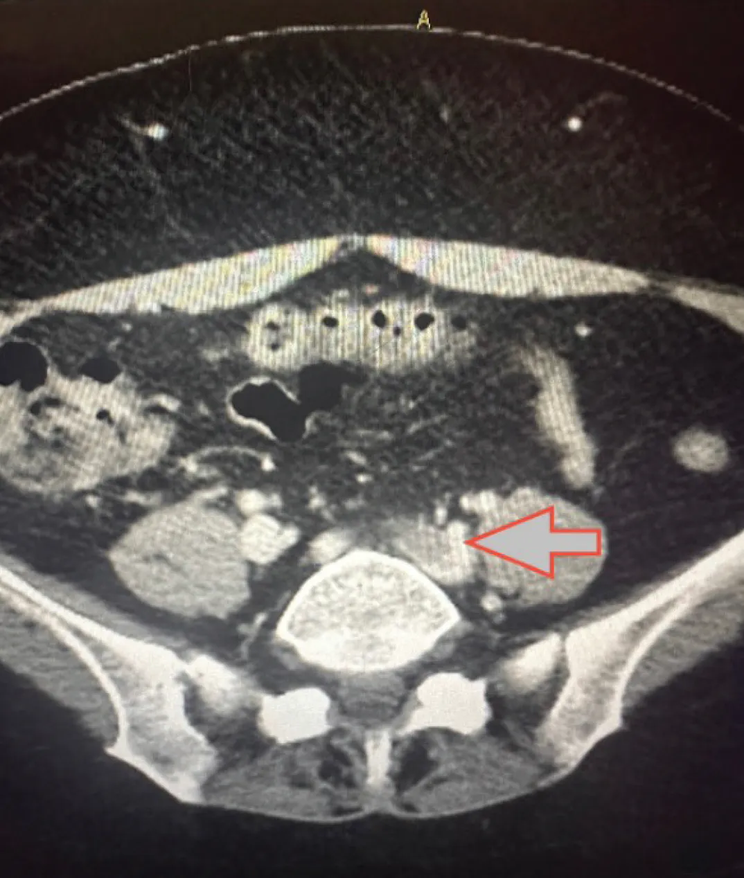
Figure 1. Axial CT of the dilated left CIA.
Vascular surgery was consulted due to involvement of the iliofemoral segment. Her right lower extremity was visibly edematous and had cyanotic congestive discoloration. She continued to be in pain despite starting the heparin drip. Given the extensive nature of the deep vein thrombosis (DVT), her young age, and physical examination findings, the decision was made to proceed with percutaneous mechanical thrombectomy with the 12-Fr Liberant™ thrombectomy system (Medtronic).
Thrombectomy Technique
The patient was taken to the operating room and placed in a prone position. The right popliteal space was prepped and draped, and the below-knee popliteal vein was accessed. An 8-Fr, 10-cm Pinnacle™* sheath (Terumo Interventional Systems) was introduced via the Seldinger technique. This was later upsized to an 11-Fr, 10-cm sheath. Diagnostic venography was performed, which demonstrated findings compatible with the venous DUS (Figure 2). Systemic heparin was readily given at 100 units/kg. The wire was advanced with a GlideCath™* catheter (Terumo Interventional Systems) to the inferior vena cava (IVC). Cavography confirmed no thrombus burden above the confluence site.
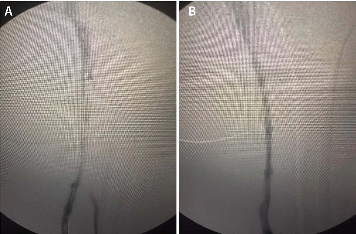
Figure 2. Fluoroscopic venogram of the left femoropopliteal vein with a guidewire (A) and catheter (B) at acute thrombus site.
I have found that the Liberant thrombectomy system works best by initiating aspiration in open flow prior to engaging the thrombus. Thus, I advanced the 12-Fr Liberant device with the inner dilator over the wire, into the IVC, and past the confluence. After the first pass, I removed the wire and was able to freely advance the catheter without difficulty. I have found that the torqueability of the Liberant catheter is one of its greatest strengths.
Given that the thrombus appeared and felt very acute, I opted not to use the clotbuster or the Excipio™* thrombectomy device (Contego Medical), which is a helpful adjunct in subacute thrombotic cases. I was able to advance the device a total of three times (back and forth) and successfully cleared out the entirety of the thrombus with an estimated blood loss of 120 mL. Follow-up venography demonstrated wide patency (Figure 3), and intravascular ultrasound (IVUS) did not reveal any need for iliac vein angioplasty/stenting. The sheath was removed from the popliteal space, and after hemostasis was achieved, a compression bandage was used from the foot to the groin. The heparin drip was readily restarted in the recovery area. The patient was transitioned to rivaroxaban and was discharged the following day.
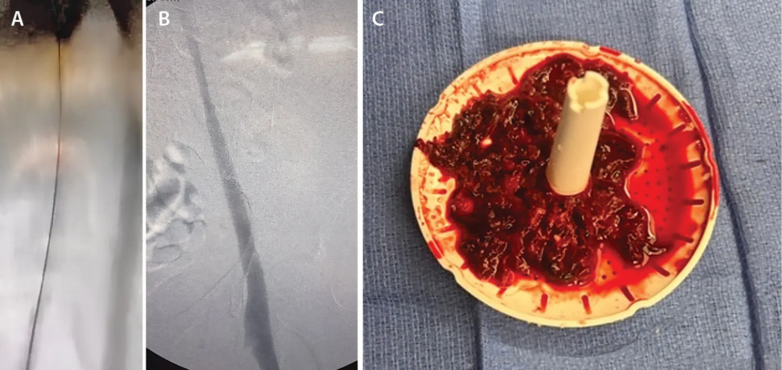
Figure 3. Follow-up fluoroscopic venogram of the femoropopliteal segment (A). Venogram demonstrating patent iliac venous flow after thrombectomy (B) and remnants of soft tissue blockage from index aspiration (C).
Postprocedure Follow-Up
I follow up with all my patients who have undergone thrombectomy after 1 month with a venous DUS. This patient’s DUS demonstrated widely patent veins apart from chronic thrombus in the proximal posterior tibial veins. The patient had no complaints and was back to pursuing her normal lifestyle.
REFLECTIONS
I believe percutaneous mechanical aspiration venous thrombectomy may play an important role in selected patients with acute iliofemoral DVT by accelerating symptom relief and functional recovery, while reducing the severity of postthrombotic syndrome and its associated long-term sequelae.1,2 It is very important to assess with IVUS to rule out underlying mechanical causes and correct them readily. I have also found it to be crucial to restart anticoagulation immediately after the intervention while the patient is in the recovery area.
1. Comerota AJ, Kearon C, Gu CS, et al. Endovascular thrombus removal for acute iliofemoral deep vein thrombosis. Circulation. 2019;139:1162-1173. doi:10.1161/CIRCULATIONAHA.118.037425
2. Vedantham S, Desai KR, Weinberg I, et. al. Society of Interventional Radiology position statement on the endovascular management of acute iliofemoral deep vein thrombosis. J Vasc Interv Radiol. 2022;34:284-299.e7. doi: 10.1016/j.jvir.2022.10.038.
Disclosures: None.
Advertisement
Advertisement